Alameda Aesthetic Dentistry
Enhancing Dentistry with
3D Imaging
Physicians have relied on computerized axial tomography scans (CAT) for many years. CAT scans are an X-ray procedure that uses many different X-ray images with the help of computers to generate cross-sectional or even 3D views of internal organs and structures within the body. A knee replacement surgery, for example, would never be performed without first examining 3D imaging.
More recently however, dentists have begun to rely on 3D imaging techniques to provide them with a detailed view of the mouth and skull. The advantage that 3D imaging holds over regular dental x-rays is that bone structure, bone density, tissues, and nerves can be viewed clearly.
3D scans can be completed in less than half a minute. This means that far less radiation enters the body than if a regular set of bitewing X-rays were taken. The main use for 3D scans is to aid to plan dental implant treatment and other oral surgery.
Dental implants are the most sophisticated replacement for missing teeth, but have historically proven to be time-consuming to place. 3D CT scans vastly reduce the time it takes to place implants. It is thought that in the near future implants will be placed in a single visit because of this unique type of imaging.
How Are 3d Scans Used?
3D scans are advantageous because they allow the dentist to magnify specific areas of the face. In addition, the dentist can easily view cross-sectional “slices” of the jaw, which makes planning treatment easier and faster.
Here are some of the main ways in which 3D scans are used in dentistry:
- Assess the quality of the jawbone where the implant will be placed.
- Determine where nerves are located.
- Diagnose tumors and disease in the early stages.
- Measure the density of the jawbone where the implant will be placed.
- Pinpoint the most effective placement for implants, including the angle of best fit.
- Plan the complete surgical procedure in advance, from start to finish.
- Precisely decide on the appropriate size and type of implants.
- View exact orientation and position of each tooth.
- View impacted teeth.
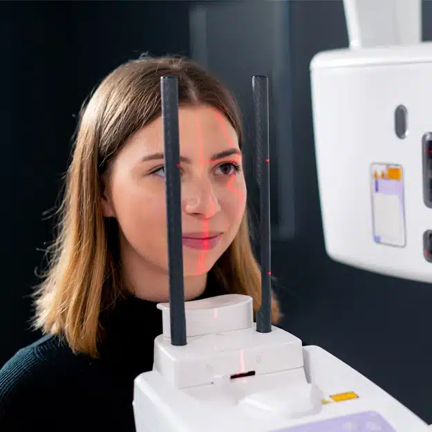
How Are 3d Scans Performed?
3D scans are quick and simple to perform. A Cone Beam Imaging System is at the heart of this type of scan. During the scan, the patient sits stationary on a designated seat. The cone beams are used to take literally hundreds of pictures of the face. These pictures are used to compile an exact 3D image of the inner mechanisms of the face and jaw. The dentist is able to zoom in on specific areas and view them from alternate angles.